Online first
Bieżący numer
Archiwum
O czasopiśmie
Polityka etyki publikacyjnej
System antyplagiatowy
Instrukcje dla Autorów
Instrukcje dla Recenzentów
Rada Redakcyjna
Komitet Redakcyjny
Recenzenci
Wszyscy recenzenci
2024
2023
2022
2021
2020
2019
2018
2017
2016
Kontakt
Bazy indeksacyjne
Klauzula przetwarzania danych osobowych (RODO)
PRACA PRZEGLĄDOWA
Wykorzystanie kleszczowych linii komórkowych w badaniach patogenów odkleszczowych
1
Zakład Biologicznych Szkodliwości Zdrowotnych i Parazytologii, Instytut Medycyny Wsi im. Witolda Chodźki, Lublin, Polska
Autor do korespondencji
Violetta Zając
Zakład Biologicznych Szkodliwości Zdrowotnych i Parazytologii, Instytut Medycyny Wsi im. Witolda Chodźki, Lublin, Polska, Jaczewskiego 2, 20-090, Lublin, Polska
Zakład Biologicznych Szkodliwości Zdrowotnych i Parazytologii, Instytut Medycyny Wsi im. Witolda Chodźki, Lublin, Polska, Jaczewskiego 2, 20-090, Lublin, Polska
Med Og Nauk Zdr. 2021;27(4):349-355

SŁOWA KLUCZOWE
DZIEDZINY
STRESZCZENIE
Wprowadzenie i cel:
Kleszcze to stawonogi odżywiające się krwią zwierząt i ludzi. Transmitują chorobotwórcze bakterie, wirusy i pierwotniaki, w tym krętkiBorrelia burgdorferi wywołujące boreliozę z Lyme i wirusa kleszczowego zapalenia mózgu. Hodowle komórkowe wyprowadzone z tkanek wek- torów znalazły szerokie zastosowanie w badaniach in vitro. Celem pracy był przegląd literatury dotyczącej kleszczowych linii komórkowych oraz ich zastosowania w badaniach naukowych.
Metody przeglądu:
W pracy opisano artykuły wyszukane w bazie PubMed na podstawie słów kluczowych: „kleszczowa linia komórkowa” (ang. tick cell line) lub „linia komórkowa z kleszczy” (ang. cell line from tick).
Opis stanu wiedzy:
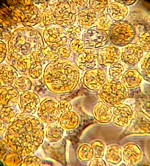
Pierwszą kleszczową linię komórkową wyprowadzoną zRhipicephalus appendiculatus opisano w 1975 roku. Obecnie wyprowadzono ponad 50 ciągłych linii komórkowych z różnych gatunków kleszczy. Większość z nich należy do embrionalnych, czyli wyprowadzonych z jaj złożonych przez samicę kleszcza. Z gatunku Ixodes ricinus, najczęściej spotykanego w Polsce, dostępne są 4 linie komórkowe. Linie są szeroko wykorzystywane w badaniach naukowych nad biologią i fizjologią kleszczy, ich odpornością na akarycydy oraz poświęconych poznaniu układu immunologicznego kleszczy. Hodowle znalazły zastosowanie w badaniach interakcji wektor–patogen, charakterystyce mikroorganizmów oraz w izolacji licznych patogennych i niepatogennych drobnoustrojów ze środowiska.
Podsumowanie:
Skuteczność zastosowania kleszczowych linii komórkowych jako narzędzia badawczego potwierdza stale rosnąca liczba prac naukowych z ich wykorzystaniem. Ich użycie może mieć znaczenie dla opracowania skutecznej diagnostyki, profilaktyki oraz w leczeniu chorób odkleszczowych. Dzięki stale rozwijającym się nowym metodom badawczym kleszczowe linie komórkowe mogą być przydatnym narzędziem w przyszłych pracach naukowych.
Kleszcze to stawonogi odżywiające się krwią zwierząt i ludzi. Transmitują chorobotwórcze bakterie, wirusy i pierwotniaki, w tym krętkiBorrelia burgdorferi wywołujące boreliozę z Lyme i wirusa kleszczowego zapalenia mózgu. Hodowle komórkowe wyprowadzone z tkanek wek- torów znalazły szerokie zastosowanie w badaniach in vitro. Celem pracy był przegląd literatury dotyczącej kleszczowych linii komórkowych oraz ich zastosowania w badaniach naukowych.
Metody przeglądu:
W pracy opisano artykuły wyszukane w bazie PubMed na podstawie słów kluczowych: „kleszczowa linia komórkowa” (ang. tick cell line) lub „linia komórkowa z kleszczy” (ang. cell line from tick).
Opis stanu wiedzy:
Pierwszą kleszczową linię komórkową wyprowadzoną zRhipicephalus appendiculatus opisano w 1975 roku. Obecnie wyprowadzono ponad 50 ciągłych linii komórkowych z różnych gatunków kleszczy. Większość z nich należy do embrionalnych, czyli wyprowadzonych z jaj złożonych przez samicę kleszcza. Z gatunku Ixodes ricinus, najczęściej spotykanego w Polsce, dostępne są 4 linie komórkowe. Linie są szeroko wykorzystywane w badaniach naukowych nad biologią i fizjologią kleszczy, ich odpornością na akarycydy oraz poświęconych poznaniu układu immunologicznego kleszczy. Hodowle znalazły zastosowanie w badaniach interakcji wektor–patogen, charakterystyce mikroorganizmów oraz w izolacji licznych patogennych i niepatogennych drobnoustrojów ze środowiska.
Podsumowanie:
Skuteczność zastosowania kleszczowych linii komórkowych jako narzędzia badawczego potwierdza stale rosnąca liczba prac naukowych z ich wykorzystaniem. Ich użycie może mieć znaczenie dla opracowania skutecznej diagnostyki, profilaktyki oraz w leczeniu chorób odkleszczowych. Dzięki stale rozwijającym się nowym metodom badawczym kleszczowe linie komórkowe mogą być przydatnym narzędziem w przyszłych pracach naukowych.
Introduction and objective:
Ticks are blood-feeding parasites of animals and humans belonging to arthropods. They transmit pathogenic bacteria, viruses, and protozoa, including Borrelia burgdorferi (causative agent of Lyme borreliosis) and tick-borne encephalitis virus. Cell cultures derived from vector tissues are tools for a wide range of in vitro studies. The aim of the study was to review the literature on tick cell lines and their application in scientific research. Review methods. The terms “tick cell line” or “cell line from tick” were used in searching the PubMed® database
Review methods:
The terms “tick cell line” or “cell line from tick” were used in searching the PubMed® database.
Brief description of the state of knowledge:
The first tick cell line derived from Rhipicephalus appendiculatus was described in 1975. Currently, more than 50 continuous cell lines from various species have been derived. Most of them are embryonic and derived from eggs laid by female ticks. Four cell lines are available from the most common tick in Poland – Ixodes ricinus. The lines are widely used in research on the biology and physiology of ticks, their resistance to acaricides, and the understanding of the immune system of ticks. Cultures have been used in the study of vector-pathogen interactions, the characteristics of microorganisms, and for the isolation of from the environment.
Summary:
The effectiveness of tick cell lines as a research tool is confirmed by an increasing number of scientific reports. Their use may be important in the development of effective diagnosis, prevention, and treatment of tick-borne diseases. Tick cell lines may be a useful tool in future research through the development of new research methods.
Sawczyn-Domańska A, Zając V. Wykorzystanie kleszczowych linii komórkowych w badaniach patogenów odkleszczowych. Med Og Nauk Zdr.
2021; 27(4): 349–355. doi: 10.26444/monz/145008
REFERENCJE (65)
1.
Černý J, Lynn G, Hrnková J, et al. Management options forIxodes ricinus-associated pathogens: a review of prevention strategies. Int J Environ Res Public Health. 2020; 17(6): 1830. https://doi.org/10.3390/ijerph....
2.
Cutler SJ, Vayssier-Taussat M, Estrada-Peña A, et al. Tick-borne diseases and co-infection: Current considerations. Ticks Tick Borne Dis. 2021; 12(1): 101607. https://doi.org/10.1016/j.ttbd....
3.
Bell-Sakyi L, Zweygarth E, Blouin EF, et al. Tick cell lines: tools for tick and tick-borne disease research. Trends Parasitol. 2007; 23(9): 450–457. https://doi.org/10.1016/j.pt.2....
4.
Varma MG, Pudney M, Leake CJ. The establishment of three cell lines from the tickRhipicephalus appendiculatus (Acari: Ixodidae) and their infection with some arboviruses. J Med Entomol. 1975; 11(6): 698–706. https://doi.org/10.1093/jmeden....
5.
Bell-Sakyi L, Darby A, Baylis M, et al. The Tick Cell Biobank: A glo - bal resource for in vitro research on ticks, other arthropods and the pathogens they transmit. Ticks Tick Borne Dis. 2018; 9(5): 1364–1371.https://doi.org/10.1016/j.ttbd....
6.
Munderloh UG, Kurtti TJ. Formulation of medium for tick cell culture. Exp. Appl. Acarol. 1989; 7: 219–229. https://doi.org/10.1007/BF0119....
7.
Bell-Sakyi L, Attoui H. Endogenous tick viruses and modulation of tick-borne pathogen growth. Front Cell Infect Microbiol. 2013; 3: 25. https://doi.org/10.3389/fcimb.....
8.
Nowak-Chmura M, Siuda K. Ticks of Poland. Review of contemporary issues and latest research. Ann Parasitol. 2012; 58(3): 125–155.
9.
Simser JA, Palmer AT, Fingerle V, et al. Rickettsia monacensis sp. nov., a spotted fever groupRickettsia, from ticks (Ixodes ricinus) collected in a European city park. Appl Environ Microbiol. 2002; 68(9): 4559–4566. https://doi.org/10.1128/AEM.68....
10.
Tick Cell Biobank. https://www.liverpool.ac.uk/li... - -research-facilities/facilities/bio-resources/tick-cell-biobank/ (access: 2021.11.08).
11.
Lim F-S, Khoo J-J, Chen F, et al. Initiation of primary cell cultures from embryonic Haemaphysalis bispinosa ticks. Syst Appl Acarol. 2017; 22(3): 323–332. http://doi.org/10.11158/saa.22....
12.
Palomar AM, Premchand-Branker S, Alberdi P, et al. Isolation of known and potentially pathogenic tick-borne microorganisms from European ixodid ticks using tick cell lines. Ticks Tick Borne Dis. 2019; 10(3): 628–638. https://doi.org/10.1016/j.ttbd....
13.
Al-Rofaai A, Bell-Sakyi L. Tick cell lines in research on tick control. Front. Physiol. 2020; 11: 152. https://doi.org/10.338 /fphys.2020.00152.
14.
Shaw DK, Wang X, Brown LJ, et al. Infection-derived lipids elicit an immune deficiency circuit in arthropods. Nat Commun. 2017; 8: 14401. https://doi.org/10.1038/ncomms....
15.
Oliva Chávez AS, Wang X, Marnin L, et al. Tick extracellular vesicles enable arthropod feeding and promote distinct outcomes of bacterial infection. Nat Commun. 2021; 12(1): 3696. https://doi.org/10.1038/ s41467-021-23900-8.
16.
Reif KE, Ujczo JK, Alperin DC, et al. Francisella tularensis novicida infection competence differs in cell lines derived from United States populations of Dermacentor andersoni and Ixodes scapularis. Sci Rep. 2018; 8(1): 12685. https://doi.org/10.1038/s41598....
17.
Goodman CL, Kang DS, Stanley D. Cell line platforms support research into arthropod immunity. Insects. 2021; 12: 738. https://doi.org/10.3390/ insects12080738.
18.
Loginov DS, Loginova YF, Dycka F, et al. Tissue-specific signatures in tick cell line MS profiles. Parasit Vectors. 2019; 12(1): 212. https://doi. org/10.1186/s13071-019-3460-5.
19.
Cramaro WJ, Hunewald OE, Bell-Sakyi L, et al. Genome scaffolding and annotation for the pathogen vector Ixodes ricinus by ultra-long single molecule sequencing. Parasit Vectors. 2017; 10(1): 71. https://doi. org/10.1186/s13071-017-2008-9.
20.
Al-Khafaji AM, Bell-Sakyi L, Fracasso G, et al. Isolation ofCandidatus Rickettsia vini from Belgian Ixodes arboricola ticks and propagation in tick cell lines. Ticks Tick Borne Dis. 2020; 11(6): 101511. https://doi. org/10.1016/j.ttbdis.2020.101511.
21.
Kurtti TJ, Felsheim RF, Burkhardt NY, et al.Rickettsia buchneri sp. nov., a rickettsial endosymbiont of the blacklegged tickIxodes scapularis. Int J Syst Evol Microbiol. 2015; 65(Pt 3): 965–970. https://doi.org/10.1099/ ijs.0.000047.
22.
Kholodilov IS, Litov AG, Klimentov AS, et al. Isolation and characterisation of Alongshan virus in Russia. Viruses. 2020; 12(4): 362. https://doi.org/10.3390/v12040....
23.
Najm NA, Silaghi C, Bell-Sakyi L, et al. Detection of bacteria related to Candidatus Midichloria mitochondrii in tick cell lines. Parasitol Res. 2012; 110(1): 437–442. https://doi.org/10.1007/s00436....
24.
Woldehiwet Z, Horrocks BK. Antigenicity of ovine strains ofAnaplasma phagocytophilum grown in tick cells and ovine granulocytes. J Comp Pathol. 2005; 132: 322–328. https://doi.org/10.1016/j.jcpa....
25.
Droleskey RE, Holman PJ, Craig TM, et al. Ultrastructure of Babesia bovis sexual stages as observed inBoophilus microplus cell cultures. Res Vet Sci. 1983; 34(2): 249–251.
26.
Ribeiro MF, Bastos CV, Vasconcelos MM, et al. Babesia bigemina: in vitro multiplication of sporokinetes in Ixodes scapularis (IDE8) cells. Exp Parasitol. 2009; 122(3): 192–195. https://doi.org/10.1016/j. exppara.2009.03.011.
27.
Obonyo M, Munderloh UG, Fingerle V, et al. Borrelia burgdorferi in tick cell culture modulates expression of outer surface proteins A and C in response to temperature. J Clin Microbiol. 1999; 37(7): 2137–2141. https://doi.org/10.1128/JCM.37....
28.
Bugrysheva J, Dobrikova EY, Godfrey HP, et al. Modulation ofBorrelia burgdorferi stringent response and gene expression during extracellular growth with tick cells. Infect Immun. 2002; 70(6): 3061–3067. https:// doi.org/10.1128/IAI.70.6.3061-3067.2002.
29.
Teixeira RC, Baêta BA, Ferreira JS, et al. Fluorescent membrane markers elucidate the association of Borrelia burgdorferi with tick cell lines. Braz J Med Biol Res. 2016; 49(7): e5211. https://doi.org/10.1590/1414-4....
30.
Kurtti TJ, Munderloh UG, Krueger DE, et al. Adhesion to and invasion of cultured tick (Acarina: Ixodidae) cells by Borrelia burgdorferi (Spirochaetales: Spirochaetaceae) and maintenance of infectivity. J Med Entomol. 1993; 30(3): 586–596. https://doi.org/10.1093/jmeden....
31.
Moniuszko A, Rückert C, Alberdi MP, et al. Coinfection of tick cell lines has variable effects on replication of intracellular bacterial and viral pathogens. Ticks Tick Borne Dis. 2014; 5(4): 415–422. https://doi. org/10.1016/j.ttbdis.2014.01.010.
32.
Taank V, Zhou W, Zhuang X, et al. Characterization of tick organic anion transporting polypeptides (OATPs) upon bacterial and viral infections. Parasit Vectors. 2018; 11(1): 593. https://doi.org/10.1186/ s13071-018-3160-6.
33.
Mattila JT, Munderloh UG, Kurtti TJ. Phagocytosis of the Lyme disease spirochete, Borrelia burgdorferi, by cells from the ticks,Ixodes scapularis and Dermacentor andersoni, infected with an endosymbiont,Rickettsia peacockii. J Insect Sci. 2007; 7: 58. https://doi.org/10.1673/031.00....
34.
Munderloh UG, Park YJ, Dioh JM, et al. Plasmid modifications in a tick-borne pathogen, Borrelia burgdorferi, cocultured with tick cells. Insect Mol Biol. 1993; 1(4): 195–203. https://doi.org/10.1111/j.1365.... tb00092.x.
35.
Fingerle V, Laux H, Munderloh UG, et al. Differential expression of outer surface proteins A and C by individual Borrelia burgdorferi in different genospecies. Med Microbiol Immunol. 2000; 189(2): 59–66. https://doi.org/10.1007/pl0000....
36.
Varela AS, Luttrell MP, Howerth EW, et al. First culture isolation of Borrelia lonestari , putative agent of southern tick-associated rash illness. J Clin Microbiol. 2004; 42(3): 1163–1169. https://doi.org/10.1128/ JCM.42.3.1163-1169.2004.
37.
Helmová R, Hönig V, Tykalová H, et al. Tick-borne encephalitis virus adaptation in different host environments and existence of quasispecies. Viruses. 2020; 12(8): 902. https://doi.org/10.3390/v12080....
38.
Mansfield KL, Cook C, Ellis RJ, et al. Tick-borne pathogens induce differential expression of genes promoting cell survival and host resi -stance in Ixodes ricinus cells. Parasit Vectors. 2017; 10(1): 81. https://doi.org/10.1186/s13071....
39.
Weisheit S, Villar M, Tykalová H, et al. Ixodes scapularis and Ixodes ricinus tick cell lines respond to infection with tick-borne encephalitis virus: transcriptomic and proteomic analysis. Parasit Vectors. 2015; 8: 599. https://doi.org/10.1186/s13071....
40.
Loginov DS, Böttinger K, Loginova YF, et al. Biotyping of IRE/CTVM19 tick cell line infected by tick-borne encephalitis virus. Ticks Tick Borne Dis. 2020; 11(4): 101420. https://doi.org/10.1016/j.ttbd....
41.
Belova OA, Litov AG, Kholodilov IS, et al. Properties of the tick-borne encephalitis virus population during persistent infection of ixodid ticks and tick cell lines. Ticks Tick Borne Dis. 2017; 8(6): 895–906. https:// doi.org/10.1016/j.ttbdis.2017.07.008.
42.
Růzek D, Bell-Sakyi L, Kopecký J, et al. Growth of tick-borne ence - phalitis virus (European subtype) in cell lines from vector and non-vector ticks. Virus Res. 2008; 137(1): 142–146. https://doi.org/10.1016/j. virusres.2008.05.013.
43.
Senigl F, Grubhoffer L, Kopecky J. Differences in maturation of tick -borne encephalitis virus in mammalian and tick cell line. Intervirology. 2006; 49(4): 239–248. https://doi.org/10.1159/000091....
44.
Lattová E, Straková P, Pokorná-Formanová P, et al. Comprehensive N-glycosylation mapping of envelope glycoprotein from tick-borne encephalitis virus grown in human and tick cells. Sci Rep. 2020; 10(1): 13204. https://doi.org/10.1038/s41598....
45.
Dyachenko V, Geiger C, Pantchev N, et al. Isolation of canineAnaplasma phagocytophilum strains from clinical blood samples using the Ixodes ricinus cell line IRE/CTVM20. Vet Microbiol. 2013; 162(2–4): 980–986. https://doi.org/10.1016/j.vetm....
46.
Passos LM. In vitro cultivation ofAnaplasma marginale and A. phagocytophilum in tick cell lines: a review. Rev Bras Parasitol Vet. 2012; 21(2):81–86. https://doi.org/10.1590/s1984-....
47.
Massung RF, Levin ML, Munderloh UG, et al. Isolation and propagation of the Ap-Variant 1 strain ofAnaplasma phagocytophilum in a tick cell line. J Clin Microbiol. 2007; 45(7): 2138–2143. https://doi.org/10.1128/ JCM.00478-07.
48.
Massung RF, Levin ML, Munderloh UG, et al. Isolation ofAnaplasma phagocytophilum strain Ap-variant 1 in a tick-derived cell line. Ann NY Acad Sci. 2006; 1078: 541–544. https://doi.org/10.1196/annals....
49.
Silaghi C, Kauffmann M, Passos LM, et al. Isolation, propagation and preliminary characterisation ofAnaplasma phagocytophilum from roedeer (Capreolus capreolus) in the tick cell line IDE8. Ticks Tick Borne Dis. 2011; 2(4): 204–208. https://doi.org/10.1016/j.ttbd....
50.
Cabezas-Cruz A, Alberdi P, Valdés JJ, et al.Anaplasma phagocytophilum infection subverts carbohydrate metabolic pathways in the tick vector, Ixodes scapularis. Front Cell Infect Microbiol. 2017a; 7: 23. https://doi. org/10.3389/fcimb.2017.00023.
51.
Cabezas-Cruz A, Alberdi P, Valdes JJ, et al. Remodeling of tick cy - toskeleton in response to infection with Anaplasma phagocytop-hilum. Front Biosci (Landmark Ed). 2017b; 22: 1830–1844. https://doi. org/10.2741/4574.
52.
Cabezas-Cruz A, Alberdi P, Ayllón N, et al. Anaplasma phagocytop- hilum increases the levels of histone modifying enzymes to inhibit cell apoptosis and facilitate pathogen infection in the tick vector Ixodes scapularis. Epigenetics. 2016; 11(4): 303–319. https://doi.org/10.1080/155922....
53.
Cabezas-Cruz A, Espinosa PJ, Obregón DA, et al. Ixodes scapularis tick cells control Anaplasma phagocytophilum infection by increasing the synthesis of phosphoenolpyruvate from tyrosine. Front Cell Infect Microbiol. 2017c; 7: 375. https://doi.org/10.3389/fcimb.....
54.
Alberdi P, Mansfield KL, Manzano-Román R, et al. Tissue-specific sig- natures in the transcriptional response toAnaplasma phagocytophilum infection of Ixodes scapularis and Ixodes ricinus tick cell lines. Front Cell Infect Microbiol. 2016; 6: 20. https://doi.org/10.3389/fcimb.....
55.
Alberdi P, Ayllón N, Cabezas-Cruz A, et al. Infection of Ixodes spp. tick cells with different Anaplasma phagocytophilum isolates induces the inhibition of apoptotic cell death. Ticks Tick Borne Dis. 2015; 6(6): 758–767. https://doi.org/10.1016/j.ttbd....
56.
Ayllón N, Villar M, Busby AT, et al. Anaplasma phagocytophilum inhibits apoptosis and promotes cytoskeleton rearrangement for in fection of tick cells. Infect Immun. 2013; 81(7): 2415–2425. https://doi. org/10.1128/IAI.00194-13.
57.
Crosby FL, Munderloh UG, Nelson CM, et al. Disruption of VirB6 paralogs in Anaplasma phagocytophilum attenuates its growth. J Bacteriol. 2020; 202(23): e00301–20. https://doi.org/10.1128/JB.003....
58.
Mateos-Hernández L, Pipová N, Allain E, et al. Enlisting the Ixodes scapularis embryonic ISE6 cell line to investigate the neuronal basis of tick-pathogen interactions. Pathogens. 2021; 10(1): 70. https://doi. org/10.3390/pathogens10010070.
59.
Contreras M, Alberdi P, Fernández De Mera IG, et al. Vaccinomics approach to the identification of candidate protective antigens for the control of tick vector infestations and Anaplasma phagocytophilum in- fection. Front Cell Infect Microbiol. 2017; 7: 360 https://doi.org/10.3389/ fcimb.2017.00360.
60.
Thu MJ, Qiu Y, Kataoka-Nakamura C, et al. Isolation of Rickettsia, Rickettsiella, and Spiroplasma from questing ticks in Japan using arthropod cells. Vector Borne Zoonotic Dis. 2019; 19(7): 474–485. https://doi.org/10.1089/vbz.20....
61.
Santibáñez S, Portillo A, Palomar AM, et al. Isolation and maintenance of Rickettsia raoultii in a Rhipicephalus sanguineus tick cell line. Microbes Infect. 2015; 17(11–12): 866–869. https://doi.org/10.1016/j. micinf.2015.09.018.
62.
Alberdi MP, Nijhof AM, Jongejan F, et al. Tick cell culture isolation and growth of Rickettsia raoultii from Dutch Dermacentor reticulatus ticks. Ticks Tick Borne Dis. 2012; 3(5–6): 349–354. https://doi.org/10.1016/j.ttbd....
63.
Husin NA, Khoo JJ, Zulkifli MMS, et al. Replication kinetics of Rickettsia raoultii in Tick Cell Lines. Microorganisms. 2021; 9(7): 1370. https:// doi.org/10.3390/microorganisms9071370.
64.
Wijnveld M, Schötta AM, Pintér A, et al. Novel Rickettsia raoultii strain isolated and propagated from AustrianDermacentor reticulatus ticks. Parasit Vectors. 2016; 9(1): 567. https://doi.org/10.1186/s13071....
65.
Herrin B, Mahapatra S, Blouin EF, et al. Growth ofCoxiella burnetii in the Ixodes scapularis-derived IDE8 tick cell line. Vector Borne Zoonotic Dis. 2011; 11(7): 917–922. https://doi.org/10.1089/vbz.20....
Udostępnij
ARTYKUŁ POWIĄZANY
| eISSN: | 2084-4905 |
| ISSN: | 2083-4543 |
Przetwarzamy dane osobowe zbierane podczas odwiedzania serwisu. Realizacja funkcji pozyskiwania informacji o użytkownikach i ich zachowaniu odbywa się poprzez dobrowolnie wprowadzone w formularzach informacje oraz zapisywanie w urządzeniach końcowych plików cookies (tzw. ciasteczka). Dane, w tym pliki cookies, wykorzystywane są w celu realizacji usług, zapewnienia wygodnego korzystania ze strony oraz w celu monitorowania ruchu zgodnie z Polityką prywatności. Dane są także zbierane i przetwarzane przez narzędzie Google Analytics (więcej).
Możesz zmienić ustawienia cookies w swojej przeglądarce. Ograniczenie stosowania plików cookies w konfiguracji przeglądarki może wpłynąć na niektóre funkcjonalności dostępne na stronie.
Możesz zmienić ustawienia cookies w swojej przeglądarce. Ograniczenie stosowania plików cookies w konfiguracji przeglądarki może wpłynąć na niektóre funkcjonalności dostępne na stronie.