Online first
Bieżący numer
Archiwum
O czasopiśmie
Polityka etyki publikacyjnej
System antyplagiatowy
Instrukcje dla Autorów
Instrukcje dla Recenzentów
Rada Redakcyjna
Komitet Redakcyjny
Recenzenci
Wszyscy recenzenci
2025
2024
2023
2022
2021
2020
2019
2018
2017
2016
Kontakt
Bazy indeksacyjne
Klauzula przetwarzania danych osobowych (RODO)
PRACA PRZEGLĄDOWA
Achalazja – omówienie aktualnego stanu wiedzy
1
Pracownia Endoskopii Przewodu Pokarmowego Uniwersyteckiego Centrum Klinicznego im. prof. K. Gibińskiego Śląskiego Uniwersytetu Medycznego w Katowicach, Polska
Autor do korespondencji
Błażej Szymczuk
Pracownia Endoskopii Przewodu Pokarmowego Uniwersyteckiego Centrum Klinicznego im. prof. K. Gibińskiego Śląskiego Uniwersytetu Medycznego w Katowicach., Katowice, Polska
Pracownia Endoskopii Przewodu Pokarmowego Uniwersyteckiego Centrum Klinicznego im. prof. K. Gibińskiego Śląskiego Uniwersytetu Medycznego w Katowicach., Katowice, Polska
Med Og Nauk Zdr. 2023;29(2):89-94

SŁOWA KLUCZOWE
DZIEDZINY
STRESZCZENIE
Wprowadzenie i cel:
Achalazja jest chorobą przewlekłą, w której dochodzi do upośledzenia funkcji relaksacyjnej mięśnia gładkokomórkowego, tworzącego dolny zwieracz przełyku, oraz zaburzeń perystaltyki przełyku, co prowadzi do zalegania treści pokarmowej powyżej dolnego zwieracza przełyku. Choroba występuje u pacjentów obu płci i może ujawnić się w każdym wieku. Praca ma na celu omówienie aktualnej wiedzy dotyczącej achalazji.
Metody przeglądu:
Do przeglądu piśmiennictwa użyto bazy PubMed oraz Google Scholar. Wyszukiwano frazy w języku angielskim: „achalasia”, „achalasia treatment”, „esophageal achalasia”, „pseudo-achalasia”.
Opis stanu wiedzy:
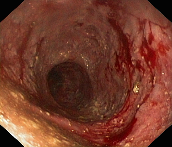
Etiologia achalazji jest złożona i obejmuje podłoże autoimmunologiczne, genetyczne, jak również infekcyjne. Najczęstszymi objawami achalazji są: regurgitacje, ból w klatce piersiowej, zgaga i przewlekły kaszel. Rozpoznanie zwykle stawiane jest po wielu latach od wystąpienia pierwszych objawów. Gruczolakorak wpustu może dawać objawy identyczne jak w przypadku achalazji (tzw. pseudoachalazja), dlatego ważne jest przeprowadzenie pełnej diagnostyki różnicowej. Leczenie achalazji jest objawowe i ma na celu ułatwienie pasażu treści pokarmowej przez przełyk oraz wpust, a co za tym idzie – poprawę jakość życia pacjenta. Wybór metody leczenia zależy od dostępności metody w danym ośrodku, doświadczenia lekarza, wyniku badania manometrycznego, stanu i preferencji pacjenta. Należy pamiętać, że pacjenci z achalazją są obarczeni zwiększonym ryzykiem rozwoju raka przełyku.
Podsumowanie:
Wczesne rozpoznanie achalazji pozwala wdrożyć odpowiednie leczenie poprawiające komfort życia oraz zmniejszające ryzyko niebezpiecznych powikłań. Etiologia achalazji nie jest do końca poznana i wymaga dalszych badań.
Achalazja jest chorobą przewlekłą, w której dochodzi do upośledzenia funkcji relaksacyjnej mięśnia gładkokomórkowego, tworzącego dolny zwieracz przełyku, oraz zaburzeń perystaltyki przełyku, co prowadzi do zalegania treści pokarmowej powyżej dolnego zwieracza przełyku. Choroba występuje u pacjentów obu płci i może ujawnić się w każdym wieku. Praca ma na celu omówienie aktualnej wiedzy dotyczącej achalazji.
Metody przeglądu:
Do przeglądu piśmiennictwa użyto bazy PubMed oraz Google Scholar. Wyszukiwano frazy w języku angielskim: „achalasia”, „achalasia treatment”, „esophageal achalasia”, „pseudo-achalasia”.
Opis stanu wiedzy:
Etiologia achalazji jest złożona i obejmuje podłoże autoimmunologiczne, genetyczne, jak również infekcyjne. Najczęstszymi objawami achalazji są: regurgitacje, ból w klatce piersiowej, zgaga i przewlekły kaszel. Rozpoznanie zwykle stawiane jest po wielu latach od wystąpienia pierwszych objawów. Gruczolakorak wpustu może dawać objawy identyczne jak w przypadku achalazji (tzw. pseudoachalazja), dlatego ważne jest przeprowadzenie pełnej diagnostyki różnicowej. Leczenie achalazji jest objawowe i ma na celu ułatwienie pasażu treści pokarmowej przez przełyk oraz wpust, a co za tym idzie – poprawę jakość życia pacjenta. Wybór metody leczenia zależy od dostępności metody w danym ośrodku, doświadczenia lekarza, wyniku badania manometrycznego, stanu i preferencji pacjenta. Należy pamiętać, że pacjenci z achalazją są obarczeni zwiększonym ryzykiem rozwoju raka przełyku.
Podsumowanie:
Wczesne rozpoznanie achalazji pozwala wdrożyć odpowiednie leczenie poprawiające komfort życia oraz zmniejszające ryzyko niebezpiecznych powikłań. Etiologia achalazji nie jest do końca poznana i wymaga dalszych badań.
Introduction and objective:
Achalasia is a chronic disease in which the relaxation function of the muscles forming the lower esophageal sphincter is impaired and the peristalsis of the esophagus is disturbed,which leads to the retention of food content. The disease affects both genders, and can occur at any age. The aim of the study is to discuss the current knowledge on achalasia.
Review methods:
The PubMed and Google Scholar databases were used for the literature review. The following phrases were searched for in English: "achalasia", "achalasia treatment", "esophageal achalasia", "pseudo-achalasia".
Brief description of the state of knowledge:
The etiology of achalasia is complex and includes autoimmune, genetic and infectious causes. The most common symptoms of achalasia are regurgitation, chest pain, heartburn and chronić cough. The diagnosis is usually made many years after the onset of the first symptoms. Adenocarcinoma of the cardia may produce symptoms identical to those of achalasia (socalled pseudoachalasia), therefore it is important to perform a full differential diagnosis. Thetreatment of achalasia is symptomatic and aims to facilitate the passage of food through the esophagus and cardia, and thus improve the patient's quality of life. The choice of the method of treatment depends on the availability of the method in a given centre, the physician’s experience, the result of the manometry test, the condition and preferences of the patient. It should be remembered that patients with achalasia are at increased risk of developing esophageal cancer.
Summary:
Early diagnosis of achalasia allows for the implementation of appropriate treatment that improves the quality of life and reduces the risk of dangerous complications. The etiology of achalasia is not fully understood and requires further research.
Szymczuk B, Milczarek J, Iwan M, Wosiewicz P, Ostrowski B. Achalazja – omówienie aktualnego stanu wiedzy. Med Og Nauk Zdr. 2023; 29(2):
89–94. doi: 10.26444/monz/166771
REFERENCJE (45)
1.
Oude Nijhuis RAB, Zaninotto G, Roman S, et al. European guidelines on achalasia: United European Gastroenterology and European Society of Neurogastroenterology and Motility recommendations. United Eu-ropean Gastroenterol J. 2020;8:13–33. doi:10.1177/2050640620903213.
2.
Savarino E, Bhatia S, Roman S, et al. Achalasia. Nat Rev Dis Primers. 2022;8:28. doi:10.1038/s41572-022-00356-8.
3.
Gong EJ. Integrated Relaxation Pressure During Swallowing: An Ever-changing Metric. J Neurogastroenterol Motil. 2021;27:151–152. doi:10.5056/jnm21033.
4.
Andolfi C, Fisichella PM. Meta-analysis of clinical outcome after treatment for achalasia based on manometric subtypes. Br J Surg. 2019;106:332–341. doi:10.1002/bjs.11049.
5.
Francis DL, Katzka DA. Achalasia: update on the disease and its treatment. Gastroenterology. 2010;139:369–74. doi:10.1053/j.gastro.2010.06.024.
6.
Gaber CE, Cotton CC, Eluri S, et al. Autoimmune and viral risk factors are associated with achalasia: A case-control study. Neurogastroenterol Motil. 2022;34(7):e14312. doi:10.1111/nmo.14312.
7.
Wu XY, Liu ZQ, Wang Y, et al. The etiology of achalasia: An immune-do -minant disease. J Dig Dis. 2021;22:126–135. doi:10.1111/1751–2980.12973.
8.
Jin H, Wang B, Zhang LL, et al. Activated Eosinophils are Present in Esophageal Muscle in Patients with Achalasia of the Esophagus. Med Sci Monit. 2018;24:2377–2383. doi:10.12659/msm.909727.
9.
Furuzawa-Carballeda J, Aguilar-León D, Gamboa-Domínguez A, et al. Achalasia--An Autoimmune Inflammatory Disease: A Cross-Sectional Study. Immunol Res. 2015;2015:729217. doi:10.1155/2015/729217.
10.
Barret M, Rouquette A, Massault PP, et al. Pseudoachalasia. Clin Res Hepatol Gastroenterol. 2018;42:99–100. doi:10.1016/j.clinre.2017.05.006.
11.
Ponds FA, van Raath MI, Mohamed SMM, et al. Diagnostic features of malignancy-associated pseudoachalasia. Aliment Pharmacol Ther. 2017;45:1449–1458. doi:10.1111/apt.14057.
12.
Tsuboi K, Hoshino M, Srinivasan A, et al. Insights gained from symptom evaluation of esophageal motility disorders: a review of 4,215 patients. Digestion. 2012;85:236–242. doi:10.1159/000336072.
13.
Vaezi MF, Richter JE. Diagnosis and management of achalasia. American College of Gastroenterology Practice Parameter Committee. Am J Gastroenterol. 1999;94:3406–3412. doi:10.1111/j.1572-0241.1999.01639.x.
14.
Smart HL, Foster PN, Evans DF, et al. Twenty four hour oesophageal acidity in achalasia before and after pneumatic dilatation. Gut. 1987;28:883–887. doi:10.1136/gut.28.7.883.
15.
Sinan H, Tatum RP, Soares RV, et al. Prevalence of respiratory symptoms in patients with achalasia. Dis Esophagus. 2011;24:224–228. doi:10.1111/j.1442-2050.2010.01126.x.
16.
Fox MR, Bredenoord AJ. Oesophageal high-resolution manometry: moving from research into clinical practice. Gut. 2008;57:405–423. doi:10.1136/gut.2007.127993.
17.
Afaneh C, Turkmany KS, Ciecierega T, et al. Esophageal Dysmotility and the Utility of Barium Swallow: An Opaque Diagnosis. Gastroenterology. 2015;148:1131–1132. doi:10.1016/S0016-5085(15)33855-5.
18.
El-Takli I, O‘Brien P, Paterson WG. Clinical diagnosis of achalasia: how reliable is the barium x-ray? Can J Gastroenterol. 2006;20:335–7. doi:10.1155/2006/193823.
19.
Blonski W, Kumar A, Feldman J, et al. Timed Barium Swallow: Diagnostic Role and Predictive Value in Untreated Achalasia, Esophagogastric Junction Outflow Obstruction, and Non-Achalasia Dysphagia. Am J Gastroenterol. 2018;113:196–203. doi:10.1038/ajg.2017.370.
20.
Carlson DA, Ravi K, Kahrilas PJ, et al. Diagnosis of Esophageal Motility Disorders: Esophageal Pressure Topography vs. Conventional Line Tracing. Am J Gastroenterol. 2015;110:967–77. doi:10.1038/ajg.2015.159.
21.
Ponds FA, Bredenoord AJ, Kessing BF, et al. Esophagogastric junction distensibility identifies achalasia subgroup with manometrically normal esophagogastric junction relaxation. Neurogastroenterol Motil. 2017;29. doi:10.1111/nmo.12908.
22.
Savarino E, di Pietro M, Bredenoord AJ, et al. Use of the Functional Lumen Imaging Probe in Clinical Esophagology. Am J Gastroenterol. 2020;115:1786–1796. doi:10.14309/ajg.0000000000000773.
23.
van Hoeij FB, Ponds FA, Smout AJ, et al. Incidence and costs of achalasia in The Netherlands. Neurogastroenterol Motil. 2018;30. doi:10.1111/nmo.13195.
24.
Pasricha PJ, Ravich WJ, Hendrix TR, et al. Treatment of achalasia with intrasphincteric injection of botulinum toxin. A pilot trial. Ann Intern Med. 1994;121(8):590–1. doi:10.7326/0003-4819-121-8-199410150-00006.
25.
Tomiyama N, Honda O, Tsubamoto M et al. Anterior mediastinal tumors: diagnostic accuracy of CT and MRI. Eur J Radiol 2009;69(2):280–8. doi:10.1016/j.ejrad.2007.10.002.
26.
Biggemann L, Uhlig J, Gliem N et al. Assessment of esophageal motility disorders by real-time MRI. Eur J Radiol 2020;132:109265. doi:10.1016/j.ejrad.2020.109265.
27.
Gaillard F, Yap J, Weerakkody Y et al. Achalasia. Reference article, Radiopaedia.org (access: 2023.05.08). doi:10.53347/rID-835.
28.
Howard PJ, Maher L, Pryde A, et al. Five year prospective study of the incidence, clinical features, and diagnosis of achalasia in Edinburgh. Gut. 1992;33:1011–1015. doi:10.1136/gut.33.8.1011.
29.
Tracey JP, Traube M. Difficulties in the diagnosis of pseudoachalasia. Am J Gastroenterol. 1994;89:2014–8.
30.
Pandolfino JE, Gawron AJ. Achalasia: A Systematic Review. JAMA. 2015;313:1841–1852. doi:10.1001/jama.2015.2996.
31.
Pomenti S, Blackett JW, Jodorkovsky D. Achalasia: Diagnosis, Management and Surveillance. Gastroenterol Clin North Am. 2021;50:721–736. doi:10.1016/j.gtc.2021.07.001.
32.
Khashab MA, Vela MF, Thosani N, et al. ASGE guideline on the management of achalasia. Gastrointest Endosc. 2020;91:213–227. doi:10.1016/j.gie.2019.04.231.
33.
Weusten BLAM, Barret M, Bredenoord AJ, et al. Endoscopic management of gastrointestinal motility disorders – part 1: European Society of Gastrointestinal Endoscopy (ESGE) Guideline. Endoscopy. 2020;52:498–515. doi:10.1055/a-1171-3174.
34.
Swanström LL. Achalasia: treatment, current status and future advances. Korean J Intern Med. 2019;34:1173–1180. doi:10.3904/kjim.2018.439.
35.
Katzka DA, Castell DO. Review article: an analysis of the efficacy, perforation rates and methods used in pneumatic dilation for achalasia. Aliment Pharmacol Ther. 2011;34(8):832–9. doi:10.1111/j.1365-2036.2011.04816.x.
36.
Vaezi MF, Pandolfino JE, Yadlapati RH, et al. ACG Clinical Guidelines: Diagnosis and Management of Achalasia. Am J Gastroenterol. 2020;115:1393–1411. doi:10.14309/ajg.0000000000000731.
37.
Andrási L, Paszt A, Simonka Z, et al. Surgical Treatment of Esophageal Achalasia in the Era of Minimally Invasive Surgery. JSLS. 2021;25:e2020.00099. doi:10.4293/JSLS.2020.00099.
38.
Lynch KL, Pandolfino JE, Howden CW, et al. Major complications of pneumatic dilation and Heller myotomy for achalasia: single-center experience and systematic review of the literature. Am J Gastroenterol. 2012;107(12):1817–25. doi:10.1038/ajg.2012.332.
39.
Richter JE, Boeckxstaens GE. Management of achalasia: surgery or pneumatic dilation. Gut. 2011;60:869–876. doi:10.1136/gut.2010.212423.
40.
Kohn GP, Dirks RC, Ansari MT, et al. SAGES guidelines for the use of peroral endoscopic myotomy (POEM) for the treatment of achalasia. Surg Endosc. 2021;35:1931–1948. doi: 10.1007/s00464-020-08282-0.
41.
Yuan X, Feng Z, Zhao Y, et al. Per-oral endoscopic dual myotomy for the treatment of achalasia. Esophagus 2021;18:941–947. doi:10.1007/s10388-021-00863-9.
42.
Erbguth FJ. Historical notes on botulism, Clostridium botulinum, botulinum toxin, and the idea of the therapeutic use of the toxin. Mov Disord. 2004;19(8):S2–6. doi:10.1002/mds.20003.
43.
Nullens S, Fockens P, Bredenoord AJ. Long-term outcomes of treatments for achalasia. Curr Opin Gastroenterol. 2021;37:408–413. doi:10.1097/MOG.0000000000000744.
44.
Lee KY, Basude D. O11 Achalasia cardia management – changing experience of a tertiary paediatric gastroenterology centre. Frontline Gastroenterology 2021;12:A8-A9. doi:10.1136/flgastro-2021-bspghan.11.
45.
Tustumi F, Bernardo WM, da Rocha JRM, et al. Esophageal achalasia: a risk factor for carcinoma. A systematic review and meta-analysis. Dis Esophagus. 2017;30:1–8. doi:10.1093/dote/dox072.
| eISSN: | 2084-4905 |
| ISSN: | 2083-4543 |
Przetwarzamy dane osobowe zbierane podczas odwiedzania serwisu. Realizacja funkcji pozyskiwania informacji o użytkownikach i ich zachowaniu odbywa się poprzez dobrowolnie wprowadzone w formularzach informacje oraz zapisywanie w urządzeniach końcowych plików cookies (tzw. ciasteczka). Dane, w tym pliki cookies, wykorzystywane są w celu realizacji usług, zapewnienia wygodnego korzystania ze strony oraz w celu monitorowania ruchu zgodnie z Polityką prywatności. Dane są także zbierane i przetwarzane przez narzędzie Google Analytics (więcej).
Możesz zmienić ustawienia cookies w swojej przeglądarce. Ograniczenie stosowania plików cookies w konfiguracji przeglądarki może wpłynąć na niektóre funkcjonalności dostępne na stronie.
Możesz zmienić ustawienia cookies w swojej przeglądarce. Ograniczenie stosowania plików cookies w konfiguracji przeglądarki może wpłynąć na niektóre funkcjonalności dostępne na stronie.