Online first
Bieżący numer
Archiwum
O czasopiśmie
Polityka etyki publikacyjnej
System antyplagiatowy
Instrukcje dla Autorów
Instrukcje dla Recenzentów
Rada Redakcyjna
Komitet Redakcyjny
Recenzenci
Wszyscy recenzenci
2025
2024
2023
2022
2021
2020
2019
2018
2017
2016
Kontakt
Bazy indeksacyjne
Klauzula przetwarzania danych osobowych (RODO)
OPIS PRZYPADKU
Krótko o wysięku opłucnowym – podróż przez opis przypadku.
1
Department of Internal Diseases, ‘LATAWIEC’ Regional Specialist Hospital, Świdnica, Poland
Autor do korespondencji
Małgorzata Leciejewska
Department of Internal Diseases, ‘LATAWIEC’ Regional Specialist Hospital, Leśna 27–29, 58-100 Świdnica, Poland
Department of Internal Diseases, ‘LATAWIEC’ Regional Specialist Hospital, Leśna 27–29, 58-100 Świdnica, Poland
Med Og Nauk Zdr. 2023;29(4):339-343

SŁOWA KLUCZOWE
DZIEDZINY
STRESZCZENIE
Wysięk opłucnowy jest częstą nieprawidłowością stwierdzaną w badaniach dodatkowych u pacjentów hospitalizowanych
na Oddziałach Chorób Wewnętrznych. Prawidłowo przeprowadzona diagnostyka pozwala ustalić przyczynę płynu,
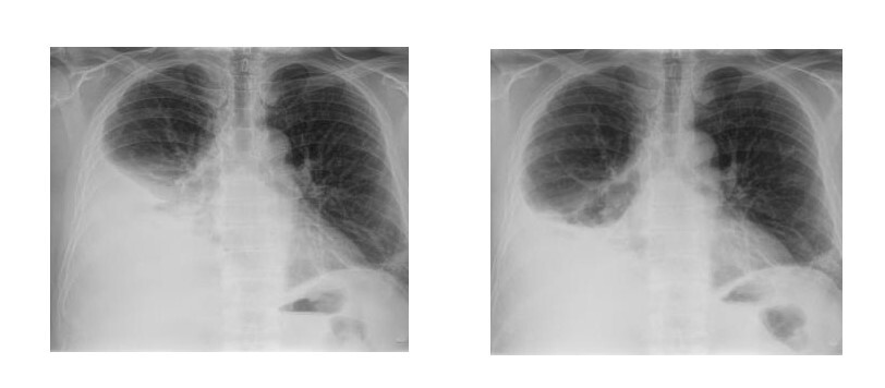
dzięki czemu możliwe jest włączenie prawidłowego leczenia. W pracy przedstawiono przypadek 63-letniego mężczyzny przyjętego do szpitala powiatowego z powodu duszności wysiłkowej. W badaniu RTG klatki piersiowej stwierdzono płyn w prawej opłucnej. Pacjenta przyjęto do Oddziału Chorób Wewnętrznych
celem diagnostyki. Ze względu na dużą ilość płynu w jamie opłucnej pacjent wymagał wielokrotnych nakłuć. W artykule omówiono najważniejsze badania diagnostyczne, jakie należy wykonać, aby ustalić przyczynę obecności płynu. Kluczową rolę przypisano rozróżnieniu etiologii płynu na podstawie kryteriów Lighta, w dalszej kolejności wzięto pod uwagę inne cechy płynu umożliwiające jego diagnostykę różnicową. Przedstawiono także możliwe punkty diagnostyczne i podjęto próbę krótkiego omówienia prawidłowego postępowania. W toku przeprowadzonej diagnostyki wysunięto podejrzenie chłoniaka opłucnej a pacjenta skierowano do ośrodka wyższej referencyjności celem dalszej diagnostyki. Przedstawiono konieczne wg autora kroki diagnostyczne konieczne do ustalenia końcowego rozpoznania. Artykuł zaprezentowano w formie pytań jednokrotnego wyboru, w których czytelnik może aktywnie uczestniczyć. Lekturę wzbogacono badaniami obrazowymi.
Pleural effusion is a common abnormality detected in additional tests in patients hospitalized in Internal Medicine Departments. Properly carried out diagnostics allows to determine the cause of the fluid, thanks to which it is possible to initiate appropriate treatment. The paper presents the case of a 63-year-old man admitted to a district hospital due to dyspnea on exertion. Chest X-ray revealed fluid in the right pleura. The patient was admitted to the Department of Internal Medicine for diagnostics. Due to the large amount of fluid in the pleural cavity, the patient required multiple punctures.
The article discusses the most important diagnostic tests that should be performed to determine the cause of the presence of fluid. A key role was assigned to distinguishing the etiology of the fluid based on Light’s criteria, and other features of the fluid enabling its differential diagnosis were taken into account. Possible diagnostic points were also presented and an attempt was made to briefly discuss the correct procedure. During the diagnostics, pleural lymphoma was suspected and the patient was referred to a higher-reference center for further diagnostics. The author presents the necessary diagnostic steps necessary to establish the final diagnosis. The article is presented in the form of single-choice questions in which the reader can actively participate. The reading was enriched with imaging studies.
Leciejewska M. Pleural effusion – a case report. J Pre-Clin Clin Res. 2023; 29(4): 333–337. doi: 10.26444/monz/176748
REFERENCJE (12)
1.
Gajewski P, editor. Interna Szczeklika 2023. Medycyna Praktyczna. 2023;II B:701–702.
2.
Antczak A, Pulmonology part 1.2nd ed. Warsaw: Medical Tribune Polska; 2020. p. 455–475.
3.
Bibby A, Dorn P, Psallidas I, Porcel J, Janssen J, Froudarakis M, Subotic D, Astoul P, Licht P, Schmid R, Scherpereel A, Rahman N, Maskell N, Cardillo G, ERS/EACTS statement on the management of malignant pleural effusions. Eur J Cardiothorac Surg. 2019;55:116–132.
4.
Ferreiro L, Toubes M, San José M, Suárez-Antelo J, Golpe A, Valdés L, Advances in pleural effusion diagnostics. Expert Review of Respir Med. 2020;14:51–66.
5.
Hooper C, Lee G, Maskell N, Investigation of a unilateral pleural effusion in adults: British Thoracic Society pleural disease guideline 2010. Thorax. 2010;65.Suppl 2:ii4-ii17.
6.
Corcoran J, et al. Pleural infection: past, present, and future directions. Lancet Respir Med. 2015;3.7:563–577.
7.
Kołodziej J, Kacprzak G. Mechanism of pleural fluid formation and diagnosis. Fam Med Prim Care Rev. 2011;4:631–639.
8.
Jany B, Welte T, Pleural Effusion in Adults-Etiology, Diagnosis, and Treatment. Dtsch Arztebl Int. 2019;116(21):377–386.
9.
Beaudoin S, Gonzalez A, Evaluation of the patient with pleural effusion CMAJ. 2018;190(10):E291-E295.
10.
Arnold D, Duneesha De Fonseka, Siobhan P, Morley A, Harvey J, Medford A, Brett M, Maskell N, Investigating unilateral pleural effusions: the role of cytology. Eur Respir J. 2018;52:1801254.
11.
Das M, Dilip K. Serous effusions in malignant lymphomas: a review. Diagn Cytopathol. 2006;34:335–347.
12.
Shimada K, Hayakawa F, Kiyoi H, Biology and management of primary effusion lymphoma. Blood. 2018;132:1879–1888.
| eISSN: | 2084-4905 |
| ISSN: | 2083-4543 |
Przetwarzamy dane osobowe zbierane podczas odwiedzania serwisu. Realizacja funkcji pozyskiwania informacji o użytkownikach i ich zachowaniu odbywa się poprzez dobrowolnie wprowadzone w formularzach informacje oraz zapisywanie w urządzeniach końcowych plików cookies (tzw. ciasteczka). Dane, w tym pliki cookies, wykorzystywane są w celu realizacji usług, zapewnienia wygodnego korzystania ze strony oraz w celu monitorowania ruchu zgodnie z Polityką prywatności. Dane są także zbierane i przetwarzane przez narzędzie Google Analytics (więcej).
Możesz zmienić ustawienia cookies w swojej przeglądarce. Ograniczenie stosowania plików cookies w konfiguracji przeglądarki może wpłynąć na niektóre funkcjonalności dostępne na stronie.
Możesz zmienić ustawienia cookies w swojej przeglądarce. Ograniczenie stosowania plików cookies w konfiguracji przeglądarki może wpłynąć na niektóre funkcjonalności dostępne na stronie.